File:Ultrasonography of a suspected malignant lymph node, with Doppler.jpg
Jump to navigation
Jump to search
No higher resolution available.
Ultrasonography_of_a_suspected_malignant_lymph_node,_with_Doppler.jpg (723 × 236 pixels, file size: 68 KB, MIME type: image/jpeg)
Summary
| Description |
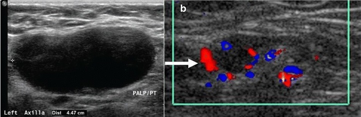
Medical ultrasonography of the axilla, showing a suspected malignant lymph node:
|
|---|---|
| Source |
Dialani, V.; James, D. F.; Slanetz, P. J. (2014). "A practical approach to imaging the axilla ". Insights into Imaging 6 (2): 217–229. doi:. ISSN 1869-4101.
|
| Date |
2014-12-23 |
| Author |
Vandana Dialani, D F James, Priscilla Slanetz |
| Permission (Reusing this file) |
See below.
|
Licensing
| This file is licensed under the Creative Commons Attribution 4.0 International license. You are free: - to share – to copy, distribute and transmit the work - to remix – to adapt the work Under the following conditions: attribution – You must attribute the work in the manner specified by the author or licensor (but not in any way that suggests that they endorse you or your use of the work). |
File history
Click on a date/time to view the file as it appeared at that time.
| Date/Time | Thumbnail | Dimensions | User | Comment | |
|---|---|---|---|---|---|
| current | 12:56, 6 February 2019 | | 723 × 236 (68 KB) | Mikael Häggström (talk | contribs) | User created page with UploadWizard |
- You cannot overwrite this file.
File usage
The following page uses this file:


