Difference between revisions of "Ultrasonography of the abdomen and pelvis"
Jump to navigation
Jump to search
m (→General screening: '') |
(→General screening: +Image) |
||
| Line 21: | Line 21: | ||
*'''Liver''', for echogenicity and focal changes. <br>''In case of suspected pathology, see: [[Ultrasonography of the liver]]'' | *'''Liver''', for echogenicity and focal changes. <br>''In case of suspected pathology, see: [[Ultrasonography of the liver]]'' | ||
*'''Biliary tract''', for gallstones (see ''[[Ultrasonography of gallstones]]'') and dilatation of the intrahepatic or extrahepatic bile ducts. The common bile duct is normally up to 8 mm.<ref name="HoeffelAzizi2006">{{cite journal|last1=Hoeffel|first1=Christine|last2=Azizi|first2=Louisa|last3=Lewin|first3=Maité|last4=Laurent|first4=Valérie|last5=Aubé|first5=Christophe|last6=Arrivé|first6=Lionel|last7=Tubiana|first7=Jean-Michel|title=Normal and Pathologic Features of the Postoperative Biliary Tract at 3D MR Cholangiopancreatography and MR Imaging|journal=RadioGraphics|volume=26|issue=6|year=2006|pages=1603–1620|issn=0271-5333|doi=10.1148/rg.266055730}}</ref> | *'''Biliary tract''', for gallstones (see ''[[Ultrasonography of gallstones]]'') and dilatation of the intrahepatic or extrahepatic bile ducts. The common bile duct is normally up to 8 mm.<ref name="HoeffelAzizi2006">{{cite journal|last1=Hoeffel|first1=Christine|last2=Azizi|first2=Louisa|last3=Lewin|first3=Maité|last4=Laurent|first4=Valérie|last5=Aubé|first5=Christophe|last6=Arrivé|first6=Lionel|last7=Tubiana|first7=Jean-Michel|title=Normal and Pathologic Features of the Postoperative Biliary Tract at 3D MR Cholangiopancreatography and MR Imaging|journal=RadioGraphics|volume=26|issue=6|year=2006|pages=1603–1620|issn=0271-5333|doi=10.1148/rg.266055730}}</ref> | ||
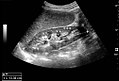
| + | [[File:Maximum length of spleen on ultrasonography.jpg|thumb|180px|Maximum length of the spleen.]] | ||
*'''Spleen''', mainly for size, where 11 cm is a common cutoff. <br>''In case of suspected pathology, see: [[Ultrasonography of the spleen]]'' | *'''Spleen''', mainly for size, where 11 cm is a common cutoff. <br>''In case of suspected pathology, see: [[Ultrasonography of the spleen]]'' | ||
Also, it is generally easy to have a quick glance at the kidneys just to exclude hydronephrosis. <br>''In case of suspected pathology, see: [[Ultrasonography of the urinary system]]'' | Also, it is generally easy to have a quick glance at the kidneys just to exclude hydronephrosis. <br>''In case of suspected pathology, see: [[Ultrasonography of the urinary system]]'' | ||
Revision as of 19:46, 11 December 2018
Author:
Mikael Häggström [notes 1]
Contents
Planning
- Choice of exam
For patients presenting with symptoms that are less specific for any certain organ, such as unspecific acute abdominal pain, consider an abdominal/pelvic CT instead.
Locations



General screening
For symptoms of the upper abdomen, it is a proper custom to perform a general screening of the following organs:
- Pancreas, mainly for dilatation of the pancreatic duct, or obvious tumors.
- Liver, for echogenicity and focal changes.
In case of suspected pathology, see: Ultrasonography of the liver - Biliary tract, for gallstones (see Ultrasonography of gallstones) and dilatation of the intrahepatic or extrahepatic bile ducts. The common bile duct is normally up to 8 mm.[1]

- Spleen, mainly for size, where 11 cm is a common cutoff.
In case of suspected pathology, see: Ultrasonography of the spleen
Also, it is generally easy to have a quick glance at the kidneys just to exclude hydronephrosis.
In case of suspected pathology, see: Ultrasonography of the urinary system
Ultrasonographies of the lower abdomen can generally be focused on the condition requested in the referral.
Diseases and conditions
Biliary tract
Appendicitis
Aneurysm
Cirrhosis
Notes
- ↑ For a full list of contributors, see article history. Creators of images are attributed at the image description pages, seen by clicking on the images. See Radlines:Authorship for details.
References
- ↑ Hoeffel, Christine; Azizi, Louisa; Lewin, Maité; Laurent, Valérie; Aubé, Christophe; Arrivé, Lionel; Tubiana, Jean-Michel (2006). "Normal and Pathologic Features of the Postoperative Biliary Tract at 3D MR Cholangiopancreatography and MR Imaging ". RadioGraphics 26 (6): 1603–1620. doi:. ISSN 0271-5333.
