Difference between revisions of "CT"
Jump to navigation
Jump to search
(+Cancer) |
|||
| (17 intermediate revisions by the same user not shown) | |||
| Line 1: | Line 1: | ||
| − | == | + | {{Top |
| − | + | |author1=[[User:Mikael Häggström|Mikael Häggström]] | |
| + | |author2= | ||
| + | }} | ||
| + | ==By location== | ||
| + | <gallery> | ||
| + | File:Computed_tomography_of_human_brain_(5)_-_local.jpg|link=CT of the head|[[CT of the head|CT of the '''head''']] | ||
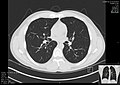
| + | File:High-resolution_computed_tomograph_of_a_normal_thorax,_axial_plane_(49).jpg|link=CT of the thorax|[[CT of the thorax|CT of the '''thorax''']] | ||
| + | File:Enlarged gallbladder with gallstone and cholecystitis.jpg|link=CT of the abdomen and pelvis|[[CT of the abdomen and pelvis|CT of the '''abdomen and pelvis''']] | ||
| + | </gallery> | ||
| + | *[[Aorta]] | ||
| + | :*[[CT of the aorta]] | ||
| + | |||
| + | ==With contrast== | ||
| + | *[[Contrast CT]] | ||
| + | |||
| + | ==General planning== | ||
| + | Upon receiving a referral for a CT, generally check: | ||
| + | *'''Who''' is the ordering individual? | ||
| + | *'''Where''': From which unit or department? | ||
| + | *'''Requested''' investigation | ||
| + | *'''Targets''': Suspected diseases or other questions or requests | ||
| + | *Patient '''history''' | ||
| + | :*Possible earlier imaging that can already answer the questions? | ||
| − | + | Consider if another modality is better for the questions at hand. | |
| − | + | ||
| − | + | Choose the proper CT settings | |
| − | + | *[[Contrast CT]] and/or non-contrast. | |
| − | + | *Include thin slices (0.5 to 1.5 mm)<ref name="MaetaniNamiki2016">{{cite journal|last1=Maetani|first1=Kazuhide|last2=Namiki|first2=Jun|last3=Matsumoto|first3=Shokei|last4=Matsunami|first4=Katsutoshi|last5=Narumi|first5=Atsushi|last6=Tsuneyoshi|first6=Toshimi|last7=Kishikawa|first7=Masanobu|title=Routine Head Computed Tomography for Patients in the Emergency Room with Trauma Requires Both Thick- and Thin-Slice Images|journal=Emergency Medicine International|volume=2016|year=2016|pages=1–4|issn=2090-2840|doi=10.1155/2016/5781790}}</ref> if the skeleton needs to be evaluated. | |
| − | |||
| − | |||
| − | |||
| − | |||
| − | |||
| − | |||
| − | |||
| − | |||
| − | |||
| − | |||
| − | |||
| − | |||
| − | |||
| − | |||
| − | |||
| − | |||
| − | |||
| − | |||
| − | |||
| − | |||
| − | |||
| − | |||
| − | |||
| − | |||
| − | |||
| − | |||
| − | |||
| − | |||
| − | |||
| − | |||
| − | |||
| − | |||
| − | |||
| − | |||
| − | |||
| − | |||
| − | |||
| − | |||
| − | |||
| − | |||
| − | |||
| − | |||
| − | |||
| − | |||
| − | |||
| − | |||
| − | |||
| − | |||
| − | |||
| − | |||
| − | |||
| − | |||
| − | |||
| − | |||
| − | |||
| − | |||
| − | |||
| − | |||
| − | |||
| − | |||
| − | |||
| − | |||
| − | |||
| − | |||
| − | |||
| − | |||
| − | |||
| − | |||
| − | |||
| − | |||
| − | |||
| − | |||
| − | | | ||
| − | | | ||
| − | | | ||
| − | | | ||
| − | | | ||
| − | | | ||
| − | | | ||
| − | | | ||
| − | |||
| − | | | ||
| − | | | ||
| − | |||
| − | |||
| − | |||
| − | |||
| − | |||
| − | |||
| − | |||
| − | |||
| − | |||
| − | |||
| − | |||
| − | |||
| − | |||
| − | |||
| − | |||
| − | |||
| − | |||
| − | |||
| − | |||
| − | |||
| − | |||
| − | |||
| − | |||
| − | |||
| − | |||
| − | |||
| − | |||
| − | |||
| − | |||
| − | |||
| − | |||
| − | |||
| − | |||
| − | |||
| − | |||
| − | |||
| − | |||
| − | + | ==General post-imaging approach== | |
| − | + | *Check the order form | |
| − | + | :*'''Who''' is the ordering individual? | |
| − | + | :*'''Where''': From which unit or department? | |
| − | + | :*Patient '''history''' | |
| − | + | :*'''Requested''' investigation | |
| − | + | :*'''Targets''': | |
| − | + | *'''Earlier''' investigations, and their findings | |
| − | + | *'''Examine''' the images at hand, including: | |
| − | + | :*'''Requested targets''' from the referral, or suspected diseases or other questions | |
| − | + | :*'''Basic screening''', whose targets vary with mainly anatomic location (see [[#Locations|locations above]]), but broadly include: | |
| − | + | ::*'''Soft tissues''', mainly for tumors or other expansions such as hematomas (see [[CT of muscle hematoma]]) | |
| − | + | ::*'''Skeleton''', mainly for fractures (see [[CT of fractures]] and tumors (see [[CT of bone tumors]]). | |
| − | + | *'''Complement''' with reconstructions where needed, such as volume renderings or thinner slices. | |
| − | + | *'''Write''' report {{Reporting}} | |
| − | + | Finally, check one extra time if the reply corresponds to the '''targets''' at hand. | |
| − | |||
| − | |||
| − | |||
| − | |||
| − | |||
| − | |||
| − | |||
| − | |||
| − | |||
| − | |||
| − | |||
| − | |||
| − | |||
| − | | | ||
| − | |||
| − | |||
| − | |||
| − | |||
| − | |||
| − | |||
| − | |||
| − | |||
| − | |||
| − | |||
| − | |||
| − | |||
| − | |||
| − | |||
| − | |||
| − | |||
| − | |||
| − | |||
| − | |||
| − | |||
| − | |||
| − | |||
| − | |||
| − | |||
| − | |||
| − | |||
| − | |||
| − | |||
| − | |||
| − | |||
| − | |||
| − | |||
| − | |||
| − | |||
| − | |||
| − | |||
| − | |||
| − | |||
| − | |||
| − | |||
| − | |||
| − | |||
| − | |||
| − | |||
| − | + | ==Hounsfield scale== | |
| − | + | *[[Hounsfield scale]] | |
| − | |||
| − | |||
| − | |||
| − | |||
| − | |||
| − | |||
| − | |||
| − | |||
| − | |||
| − | |||
| − | |||
| − | |||
| − | |||
| − | |||
| − | |||
| − | |||
| − | |||
| − | |||
| − | |||
| − | |||
| − | |||
| − | |||
| − | |||
| − | |||
| − | |||
| − | |||
| − | |||
| − | |||
| − | |||
| − | |||
| − | |||
| − | |||
| − | |||
| − | |||
| − | |||
| − | |||
| − | |||
| − | |||
| − | |||
| − | |||
| − | |||
| − | |||
| − | |||
| − | |||
| − | |||
| − | |||
| − | |||
| − | |||
| − | |||
| − | |||
| − | |||
| − | |||
| − | |||
| − | |||
| − | |||
| − | |||
| − | |||
| − | |||
| − | |||
| − | |||
| − | |||
| − | |||
| − | |||
| − | |||
| − | |||
| − | |||
| − | |||
| − | |||
| − | |||
| − | |||
| − | |||
| − | |||
| − | |||
| − | |||
| − | |||
| − | |||
| − | |||
| − | |||
| − | |||
| − | |||
| − | |||
| − | |||
| − | |||
| − | |||
| − | |||
| − | |||
| − | |||
| − | |||
| − | |||
| − | |||
| − | |||
| − | |||
| − | |||
| − | |||
| − | |||
| − | |||
| − | |||
| − | |||
| − | [[ | + | ==Location-unspecific diseases== |
| + | *[[CT of cancer|CT of '''cancer''']] | ||
| + | {{Bottom}} | ||
Latest revision as of 15:43, 25 March 2019
Author:
Mikael Häggström [notes 1]
Contents
By location


With contrast
General planning
Upon receiving a referral for a CT, generally check:
- Who is the ordering individual?
- Where: From which unit or department?
- Requested investigation
- Targets: Suspected diseases or other questions or requests
- Patient history
- Possible earlier imaging that can already answer the questions?
Consider if another modality is better for the questions at hand.
Choose the proper CT settings
- Contrast CT and/or non-contrast.
- Include thin slices (0.5 to 1.5 mm)[1] if the skeleton needs to be evaluated.
General post-imaging approach
- Check the order form
- Who is the ordering individual?
- Where: From which unit or department?
- Patient history
- Requested investigation
- Targets:
- Earlier investigations, and their findings
- Examine the images at hand, including:
- Requested targets from the referral, or suspected diseases or other questions
- Basic screening, whose targets vary with mainly anatomic location (see locations above), but broadly include:
- Soft tissues, mainly for tumors or other expansions such as hematomas (see CT of muscle hematoma)
- Skeleton, mainly for fractures (see CT of fractures and tumors (see CT of bone tumors).
- Complement with reconstructions where needed, such as volume renderings or thinner slices.
- Write report
- See also: General notes on reporting
Finally, check one extra time if the reply corresponds to the targets at hand.
Hounsfield scale
Location-unspecific diseases
Notes
- ↑ For a full list of contributors, see article history. Creators of images are attributed at the image description pages, seen by clicking on the images. See Radlines:Authorship for details.
References
- ↑ Maetani, Kazuhide; Namiki, Jun; Matsumoto, Shokei; Matsunami, Katsutoshi; Narumi, Atsushi; Tsuneyoshi, Toshimi; Kishikawa, Masanobu (2016). "Routine Head Computed Tomography for Patients in the Emergency Room with Trauma Requires Both Thick- and Thin-Slice Images ". Emergency Medicine International 2016: 1–4. doi:. ISSN 2090-2840.
