Difference between revisions of "X-ray of the thorax in tuberculosis"
(→Chest X-ray findings that can suggest active TB: Explained) |
(Choice) |
||
| (3 intermediate revisions by the same user not shown) | |||
| Line 10: | Line 10: | ||
;Indications | ;Indications | ||
Symptoms and/or previous stay in geographic area with high prevalence of tuberculosis. | Symptoms and/or previous stay in geographic area with high prevalence of tuberculosis. | ||
| − | + | {{Pulmonary tuberculosis - Choice of investigation}} | |
;Projections | ;Projections | ||
A posterior-anterior (PA) is the standard view used; other views (lateral or lordotic) or CT scans may be necessary. | A posterior-anterior (PA) is the standard view used; other views (lateral or lordotic) or CT scans may be necessary. | ||
| Line 25: | Line 25: | ||
This category comprises all findings typically associated with active pulmonary TB.<ref name=cdc/> | This category comprises all findings typically associated with active pulmonary TB.<ref name=cdc/> | ||
| − | 1. Infiltrate or consolidation - Opacification of airspaces within the lung parenchyma. Consolidation or infiltrate can be dense or patchy and might have irregular, ill-defined, or hazy borders. | + | 1. '''Infiltrate or consolidation''' - Opacification of airspaces within the lung parenchyma. Consolidation or infiltrate can be dense or patchy and might have irregular, ill-defined, or hazy borders. |
<gallery mode=packed heights=190> | <gallery mode=packed heights=190> | ||
File:Dense opacity of primary pulmonary tuberculosis.jpg|Dense homogenous opacity in right, middle and lower lobe of primary pulmonary TB. | File:Dense opacity of primary pulmonary tuberculosis.jpg|Dense homogenous opacity in right, middle and lower lobe of primary pulmonary TB. | ||
| Line 32: | Line 32: | ||
File:Chest x-ray of Ghon's complex of active tuberculosis.jpg|Chest x-ray of Ghon's complex of active tuberculosis. A Ghon's complex consists of a calcified focus of infection and an associated lymph node. | File:Chest x-ray of Ghon's complex of active tuberculosis.jpg|Chest x-ray of Ghon's complex of active tuberculosis. A Ghon's complex consists of a calcified focus of infection and an associated lymph node. | ||
</gallery> | </gallery> | ||
| − | 2. Any cavitary lesion - Lucency (darkened area) within the lung parenchyma, with or without irregular margins that might be surrounded by an area of airspace consolidation or infiltrates, or by nodular or fibrotic (reticular) densities, or both. The walls surrounding the lucent area can be thick or thin. Calcification can exist around a cavity. | + | 2. '''Any cavitary lesion''' - Lucency (darkened area) within the lung parenchyma, with or without irregular margins that might be surrounded by an area of airspace consolidation or infiltrates, or by nodular or fibrotic (reticular) densities, or both. The walls surrounding the lucent area can be thick or thin. Calcification can exist around a cavity. |
<gallery mode=packed heights=190> | <gallery mode=packed heights=190> | ||
File:Tuberculosis-x-ray-1.jpg|Chest X-ray of a person with advanced tuberculosis: Infection in both lungs is marked by white arrow-heads, and the formation of a cavity is marked by black arrows. | File:Tuberculosis-x-ray-1.jpg|Chest X-ray of a person with advanced tuberculosis: Infection in both lungs is marked by white arrow-heads, and the formation of a cavity is marked by black arrows. | ||
</gallery> | </gallery> | ||
| − | 3. Nodule with poorly defined margins - Round density within the lung parenchyma, also called a tuberculoma. Nodules included in this category are those with margins that are indistinct or poorly defined ([[tree-in-bud sign]]<ref>{{cite journal|last=Rossi|first=S. E.|author2=Franquet, T. |author3=Volpacchio, M. |author4=Gimenez, A. |author5= Aguilar, G. |title=Tree-in-Bud Pattern at Thin-Section CT of the Lungs: Radiologic-Pathologic Overview|journal=Radiographics|date=1 May 2005|volume=25|issue=3|pages=789–801|doi=10.1148/rg.253045115|url=http://radiographics.rsna.org/content/25/3/789.full|accessdate=28 May 2012}}</ref>). The surrounding haziness can be either subtle or readily apparent and suggests coexisting airspace consolidation. | + | 3. '''Nodule with poorly defined margins''' - Round density within the lung parenchyma, also called a tuberculoma. Nodules included in this category are those with margins that are indistinct or poorly defined ([[tree-in-bud sign]]<ref>{{cite journal|last=Rossi|first=S. E.|author2=Franquet, T. |author3=Volpacchio, M. |author4=Gimenez, A. |author5= Aguilar, G. |title=Tree-in-Bud Pattern at Thin-Section CT of the Lungs: Radiologic-Pathologic Overview|journal=Radiographics|date=1 May 2005|volume=25|issue=3|pages=789–801|doi=10.1148/rg.253045115|url=http://radiographics.rsna.org/content/25/3/789.full|accessdate=28 May 2012}}</ref>). The surrounding haziness can be either subtle or readily apparent and suggests coexisting airspace consolidation. |
<gallery mode=packed heights=190> | <gallery mode=packed heights=190> | ||
File:Chest X-ray and CT of tree-in-bud sign post-primary pulmonary tuberculosis.jpg|Chest x-ray showing nodule with margins that are indistinct or poorly defined (tree-in-bud sign) in post-primary pulmonary TB. | File:Chest X-ray and CT of tree-in-bud sign post-primary pulmonary tuberculosis.jpg|Chest x-ray showing nodule with margins that are indistinct or poorly defined (tree-in-bud sign) in post-primary pulmonary TB. | ||
</gallery> | </gallery> | ||
| − | + | 4. '''Pleural effusion''' - Presence of a significant amount of fluid within the pleural space. This finding must be distinguished from blunting of the costophrenic angle, which may or may not represent a small amount of fluid within the pleural space (except in children when even minor blunting must be considered a finding that can suggest active TB). | |
<gallery mode=packed heights=190>> | <gallery mode=packed heights=190>> | ||
File:Pleural effusion of primary pulmonary tuberculosis.jpg|Chest x-ray showing dense opacity pleural effusion in the lower left lung of primary pulmonary TB. | File:Pleural effusion of primary pulmonary tuberculosis.jpg|Chest x-ray showing dense opacity pleural effusion in the lower left lung of primary pulmonary TB. | ||
</gallery> | </gallery> | ||
| − | 5. Hilar or mediastinal lymphadenopathy ([[bihilar lymphadenopathy]]) - Enlargement of lymph nodes in one or both hila or within the mediastinum, with or without associated atelectasis or consolidation. | + | 5. '''Hilar or mediastinal lymphadenopathy''' ([[bihilar lymphadenopathy]]) - Enlargement of lymph nodes in one or both hila or within the mediastinum, with or without associated atelectasis or consolidation. |
<gallery mode=packed heights=190>> | <gallery mode=packed heights=190>> | ||
File:Chest x-ray of bilateral hilar adenopathy of primary pulmonary tuberculosis.jpg|Chest x-ray showing bilateral hilar adenopathy of primary pulmonary TB | File:Chest x-ray of bilateral hilar adenopathy of primary pulmonary tuberculosis.jpg|Chest x-ray showing bilateral hilar adenopathy of primary pulmonary TB | ||
</gallery> | </gallery> | ||
| − | 6. Linear, interstitial disease (in children only) - Prominence of linear, interstitial (septal) markings. | + | 6. '''Linear, interstitial disease''' (in children only) - Prominence of linear, interstitial (septal) markings. |
<gallery mode=packed heights=190>> | <gallery mode=packed heights=190>> | ||
File:Kerley B lines of primary tuberculosis.jpg|Chest x-ray showing Kerely B line due to interstitial oedema (in children only) of primary pulmonary tuberculosis. | File:Kerley B lines of primary tuberculosis.jpg|Chest x-ray showing Kerely B line due to interstitial oedema (in children only) of primary pulmonary tuberculosis. | ||
</gallery> | </gallery> | ||
| − | 7. | + | 7. '''Miliary nodules ''' - nodules of millet size (1 to 2 millimeters) distributed throughout the parenchyma. |
<gallery mode=packed heights=190> | <gallery mode=packed heights=190> | ||
| − | File: | + | File:Chest radiograph of miliary tuberculosis 1.jpg|Miliary tuberculosis, 13 days after onset, showing bilateral interstitial infiltrates |
| + | File:Chest radiograph of miliary tuberculosis 2.jpg|Miliary tuberculosis, 22 days after onset, showing extensive bilateral reticulo-nodular infiltrates | ||
</gallery> | </gallery> | ||
| Line 61: | Line 62: | ||
The main chest X-ray findings that can suggest inactive TB are:<ref name=cdc/> | The main chest X-ray findings that can suggest inactive TB are:<ref name=cdc/> | ||
| − | 1. Discrete fibrotic scar or linear | + | <br>1. '''Discrete fibrotic scar or linear opacity''' — Discrete linear or reticular densities within the lung. The edges of these densities should be distinct and there should be no suggestion of airspace opacification or haziness between or surrounding these densities. Calcification can be present within the lesion and then the lesion is called a “fibrocalcific” scar. |
<gallery mode=packed heights=190> | <gallery mode=packed heights=190> | ||
File:Chest X-ray of fibrocalcific scar after secondary tuberculosis.jpg|Chest x-ray showing fibrocalcific scar after secondary tuberculosis as air-space opacification or haziness between or surrounding these densities. | File:Chest X-ray of fibrocalcific scar after secondary tuberculosis.jpg|Chest x-ray showing fibrocalcific scar after secondary tuberculosis as air-space opacification or haziness between or surrounding these densities. | ||
</gallery> | </gallery> | ||
| − | 2. Discrete nodule(s) without | + | 2. '''Discrete nodule(s) without calcification''' - One or more nodular densities with distinct borders and without any surrounding airspace opacification. Nodules are generally round or have rounded edges. These features allow them to be distinguished from infiltrates or airspace opacities. To be included here, these nodules must be noncalcified. Nodules that are calcified are included in the category “OTHER X-ray findings, No follow-up needed”. |
<gallery mode=packed heights=190> | <gallery mode=packed heights=190> | ||
File:Chest X-ray of discrete round nodules after secondary tuberculosis.jpg|Chest x-ray showing discrete round nodule(s) with round edges without calcification, after secondary tuberculosis. | File:Chest X-ray of discrete round nodules after secondary tuberculosis.jpg|Chest x-ray showing discrete round nodule(s) with round edges without calcification, after secondary tuberculosis. | ||
</gallery> | </gallery> | ||
| − | 3. Discrete fibrotic scar with volume loss or | + | 3. '''Discrete fibrotic scar with volume loss or retraction''' — Discrete linear densities with reduction in the space occupied by the upper lobe. Associated signs include upward deviation of the fissure or hilum on the corresponding side with asymmetry of the volumes of the two thoracic cavities. |
<gallery mode=packed heights=190> | <gallery mode=packed heights=190> | ||
File:Chest x-ray of distinct fibrotic scar after secondary tuberculosis.jpg|Chest x-ray showing distinct fibrotic scar with volume loss or retraction with an upward deviation of the fissure or hilum on the corresponding side with asymmetry of the volumes of the two thoracic cavities. | File:Chest x-ray of distinct fibrotic scar after secondary tuberculosis.jpg|Chest x-ray showing distinct fibrotic scar with volume loss or retraction with an upward deviation of the fissure or hilum on the corresponding side with asymmetry of the volumes of the two thoracic cavities. | ||
</gallery> | </gallery> | ||
| − | 4. Discrete nodule(s) with volume loss or | + | 4. '''Discrete nodule(s) with volume loss or retraction''' — One or more nodular densities with distinct borders and no surrounding airspace opacification with reduction in the space occupied by the upper lobe. Nodules are generally round or have rounded edges. |
<gallery mode=packed heights=190> | <gallery mode=packed heights=190> | ||
File:Chest x-ray of discrete nodule and volume loss.jpg|Chest x-ray showing nodular densities with distinct borders and no surrounding airspace opacification, with a reduction in the space occupied by the upper lobe. | File:Chest x-ray of discrete nodule and volume loss.jpg|Chest x-ray showing nodular densities with distinct borders and no surrounding airspace opacification, with a reduction in the space occupied by the upper lobe. | ||
</gallery> | </gallery> | ||
| − | 5. | + | 5. '''Upper lobe bronchiectasis''' - Bronchial dilation with bronchial wall thickening. |
<gallery mode=packed heights=190> | <gallery mode=packed heights=190> | ||
File:Chest x-ray of bronchiectasis post-primary pulmonary tuberculosis.jpg|Chest x-ray showing course bronchiectasis of the lungs post-primary pulmonary tuberculosis. | File:Chest x-ray of bronchiectasis post-primary pulmonary tuberculosis.jpg|Chest x-ray showing course bronchiectasis of the lungs post-primary pulmonary tuberculosis. | ||
Latest revision as of 13:58, 6 May 2019
Author:
Mikael Häggström [notes 1]

Projectional radiography ("X-ray") of the thorax in suspected tuberculosis:
Contents
Planning
- Indications
Symptoms and/or previous stay in geographic area with high prevalence of tuberculosis.
Choice of investigation
For suspected pulmonary tuberculosis:
- X-ray of the thorax in tuberculosis is generally the initial choice of investigation.[1]
- CT of the thorax is feasible if:
- Projections
A posterior-anterior (PA) is the standard view used; other views (lateral or lordotic) or CT scans may be necessary.
Evaluation
The chest X-ray and classification worksheet by the Centers for Disease Control and Prevention (CDC) of the United States is designed to group findings into categories based on their likelihood of being related to TB or non-TB conditions needing medical follow-up.[3]
Normal findings
These are films that are completely normal, with no identifiable cardiothoracic or musculoskeletal abnormality.
Abnormal findings
Chest X-ray findings that can suggest active TB
This category comprises all findings typically associated with active pulmonary TB.[3]
1. Infiltrate or consolidation - Opacification of airspaces within the lung parenchyma. Consolidation or infiltrate can be dense or patchy and might have irregular, ill-defined, or hazy borders.

Dense homogenous opacity in right, middle and lower lobe of primary pulmonary TB.

Chest x-ray showing patchy opacification on the upper right and mid-zone lung with fibrotic shadows, as well as bilateral hilar lymphadenopathy.

Chest x-ray showing coarse reticulonodular densities on the lower right lung of post-primary pulmonary TB.
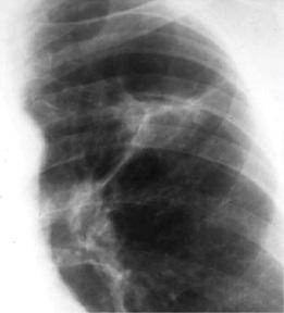
Chest x-ray of Ghon's complex of active tuberculosis. A Ghon's complex consists of a calcified focus of infection and an associated lymph node.


2. Any cavitary lesion - Lucency (darkened area) within the lung parenchyma, with or without irregular margins that might be surrounded by an area of airspace consolidation or infiltrates, or by nodular or fibrotic (reticular) densities, or both. The walls surrounding the lucent area can be thick or thin. Calcification can exist around a cavity.

Chest X-ray of a person with advanced tuberculosis: Infection in both lungs is marked by white arrow-heads, and the formation of a cavity is marked by black arrows.

3. Nodule with poorly defined margins - Round density within the lung parenchyma, also called a tuberculoma. Nodules included in this category are those with margins that are indistinct or poorly defined (tree-in-bud sign[4]). The surrounding haziness can be either subtle or readily apparent and suggests coexisting airspace consolidation.

Chest x-ray showing nodule with margins that are indistinct or poorly defined (tree-in-bud sign) in post-primary pulmonary TB.

4. Pleural effusion - Presence of a significant amount of fluid within the pleural space. This finding must be distinguished from blunting of the costophrenic angle, which may or may not represent a small amount of fluid within the pleural space (except in children when even minor blunting must be considered a finding that can suggest active TB).

Chest x-ray showing dense opacity pleural effusion in the lower left lung of primary pulmonary TB.

5. Hilar or mediastinal lymphadenopathy (bihilar lymphadenopathy) - Enlargement of lymph nodes in one or both hila or within the mediastinum, with or without associated atelectasis or consolidation.

Chest x-ray showing bilateral hilar adenopathy of primary pulmonary TB

6. Linear, interstitial disease (in children only) - Prominence of linear, interstitial (septal) markings.

Chest x-ray showing Kerely B line due to interstitial oedema (in children only) of primary pulmonary tuberculosis.

7. Miliary nodules - nodules of millet size (1 to 2 millimeters) distributed throughout the parenchyma.

Miliary tuberculosis, 13 days after onset, showing bilateral interstitial infiltrates

Miliary tuberculosis, 22 days after onset, showing extensive bilateral reticulo-nodular infiltrates


Chest X-ray findings that can suggest inactive TB
This category includes findings that are suggestive of prior TB, that is inactive. It must be remembered that assessments of the activity of TB cannot be made accurately on the basis of a single radiograph alone. If there is any question of active TB, sputum smears must be obtained. Therefore, any applicant might have findings grouped in this category, but still have active TB as suggested by the presence of signs or symptoms of TB, or sputum smears positive for AFB.[3]
The main chest X-ray findings that can suggest inactive TB are:[3]
1. Discrete fibrotic scar or linear opacity — Discrete linear or reticular densities within the lung. The edges of these densities should be distinct and there should be no suggestion of airspace opacification or haziness between or surrounding these densities. Calcification can be present within the lesion and then the lesion is called a “fibrocalcific” scar.

Chest x-ray showing fibrocalcific scar after secondary tuberculosis as air-space opacification or haziness between or surrounding these densities.

2. Discrete nodule(s) without calcification - One or more nodular densities with distinct borders and without any surrounding airspace opacification. Nodules are generally round or have rounded edges. These features allow them to be distinguished from infiltrates or airspace opacities. To be included here, these nodules must be noncalcified. Nodules that are calcified are included in the category “OTHER X-ray findings, No follow-up needed”.
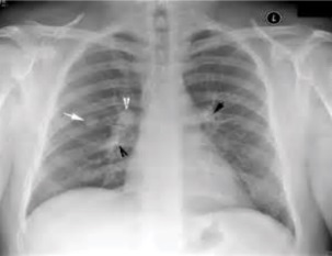
Chest x-ray showing discrete round nodule(s) with round edges without calcification, after secondary tuberculosis.

3. Discrete fibrotic scar with volume loss or retraction — Discrete linear densities with reduction in the space occupied by the upper lobe. Associated signs include upward deviation of the fissure or hilum on the corresponding side with asymmetry of the volumes of the two thoracic cavities.

Chest x-ray showing distinct fibrotic scar with volume loss or retraction with an upward deviation of the fissure or hilum on the corresponding side with asymmetry of the volumes of the two thoracic cavities.

4. Discrete nodule(s) with volume loss or retraction — One or more nodular densities with distinct borders and no surrounding airspace opacification with reduction in the space occupied by the upper lobe. Nodules are generally round or have rounded edges.

Chest x-ray showing nodular densities with distinct borders and no surrounding airspace opacification, with a reduction in the space occupied by the upper lobe.

5. Upper lobe bronchiectasis - Bronchial dilation with bronchial wall thickening.

Chest x-ray showing course bronchiectasis of the lungs post-primary pulmonary tuberculosis.

Other chest X-ray findings
Follow-up needed
This category includes findings that suggest the need for a follow-up evaluation for non-TB conditions.[3]
- Musculoskeletal abnormalities - New bony fractures or radiographically apparent bony abnormalities that need follow-up.
- Cardiac abnormalities - Cardiac enlargement or anomalies, vascular abnormalities, or any other radiographically apparent cardiovascular abnormality of significant nature to require follow-up.
- Pulmonary abnormalities - Pulmonary finding of a non-TB nature, such as a mass, that needs follow-up.
- Other - Any other finding that the panel physician believes needs follow-up, but is not one of the above.
No follow-up needed
This category includes findings that are minor and not suggestive of TB disease. These findings require no follow-up evaluation.[3].

- Pleural thickening - Irregularity or abnormal prominence of the pleural margin, including apical capping (thickening of the pleura in the apical region). Pleural thickening can be calcified.
- Diaphragmatic tenting - A localized accentuation of the normal convexity of the hemidiaphragm as if “pulled upwards by a string.”
- Blunting of costophrenic angle (in adults)—Loss of sharpness of one or both costophrenic angles. Blunting can be related to a small amount of fluid in the pleural space or to pleural thickening and, by itself, is a non-specific finding (except in children, when even minor blunting may suggest active TB). In contrast a large pleural effusion, or the presence of a significant amount of fluid in the pleural space, may be a sign of active TB at any age.
- Solitary calcified nodules or granuloma - Discrete calcified nodule or granuloma, or calcified lymph node. The calcified nodule can be within the lung, hila, or mediastinum. The borders must be sharp, distinct, and well defined. This was considered a Class B3 TB in the past; however, Class B3 has been omitted from the classification scheme because it has not been found to be associated with active TB.
- Minor musculoskeletal findings - Minor findings needing no follow-up.
- Minor cardiac findings - Minor findings needing no follow-up.
Notes
- ↑ For a full list of contributors, see article history. Creators of images are attributed at the image description pages, seen by clicking on the images. See Radlines:Authorship for details.
References
- ↑ 1.0 1.1 Bomanji, J. B.; Gupta, N.; Gulati, P.; Das, C. J. (2015). "Imaging in Tuberculosis ". Cold Spring Harbor Perspectives in Medicine 5 (6): a017814–a017814. doi:. ISSN 2157-1422.
- ↑ Alkabab, Yosra; Enani, Mushira; Indarkiri, Nouf; Heysell, Scott (2018). "Performance of computed tomography versus chest radiography in patients with pulmonary tuberculosis with and without diabetes at a tertiary hospital in Riyadh, Saudi Arabia ". Infection and Drug Resistance Volume 11: 37–43. doi:. ISSN 1178-6973.
- ↑ 3.0 3.1 3.2 3.3 3.4 3.5 . Instructions to Panel Physicians for Completing New U.S. Department of State MEDICAL EXAMINATION FOR IMMIGRANT OR REFUGEE APPLICANT (DS-2053) and Associated WORKSHEETS (DS-3024, DS-3025, and DS-3026). Centers for Disease Control and Prevention.
- ↑ Rossi, S. E.; Franquet, T.; Volpacchio, M.; Gimenez, A.; Aguilar, G. (1 May 2005). "Tree-in-Bud Pattern at Thin-Section CT of the Lungs: Radiologic-Pathologic Overview ". Radiographics 25 (3): 789–801. doi:. Archived from the original. . Retrieved on 28 May 2012.
