Difference between revisions of "Thorax"
Jump to navigation
Jump to search
(+Choice of investigation) |
(Templates) |
||
| Line 1: | Line 1: | ||
| + | {{Top | ||
| + | |author1=[[User:Mikael Häggström|Mikael Häggström]] | ||
| + | |author2= | ||
| + | }} | ||
==Modalities== | ==Modalities== | ||
<gallery> | <gallery> | ||
| Line 27: | Line 31: | ||
Superficial soft tissue diseases: | Superficial soft tissue diseases: | ||
{{Superficial soft tissue diseases}} | {{Superficial soft tissue diseases}} | ||
| + | |||
| + | {{Bottom}} | ||
Latest revision as of 11:17, 4 April 2019
Author:
Mikael Häggström [notes 1]
Contents
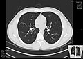
Modalities

Other modalities
- Fluoroscopy of the thorax
- Ultrasonography of the thorax
Choice of investigation
- X-ray of the thorax is generally the initial investigation of choice in dyspnea.
- CT of the thorax is generally the investigation of choice in pulmonary embolism and aortic dissection. It is recommended as complementary to X-ray in chest trauma.[1]
Locations
- Ribs
- Sternoclavicular joint
Diseases and conditions
Superficial soft tissue diseases:
Notes
- ↑ For a full list of contributors, see article history. Creators of images are attributed at the image description pages, seen by clicking on the images. See Radlines:Authorship for details.
References
- ↑ . Blunt Chest Trauma, ACR Appropriateness Criteria. American College of Radiology. Date of origin: 2013
